Commentary on the latest advances and controversies in kidney disease: Dr. Seuss 'Oh the places we will go.'
Showing posts with label Quiz. Show all posts
Thursday, September 12, 2013
Monday, August 26, 2013
Tuesday, December 11, 2012
Image Quiz - Membranous Glomerulopathy
The EM shows numerous scattered subepithelial deposits with some of the deposits were separated from each other by basement membrane spikes. There are no intramembranous and subendothelial deposits. There is extensive effacement of the foot processes of the visceral epithelial cells.
The diagnosis is membranous glomerulopathy. Here is a great reference in CJASN by Fervenza et al.
What does the EM image show? What's the diagnosis?
The diagnosis is membranous glomerulopathy. Here is a great reference in CJASN by Fervenza et al.
What does the EM image show? What's the diagnosis?
Saturday, December 8, 2012
Monday, December 3, 2012
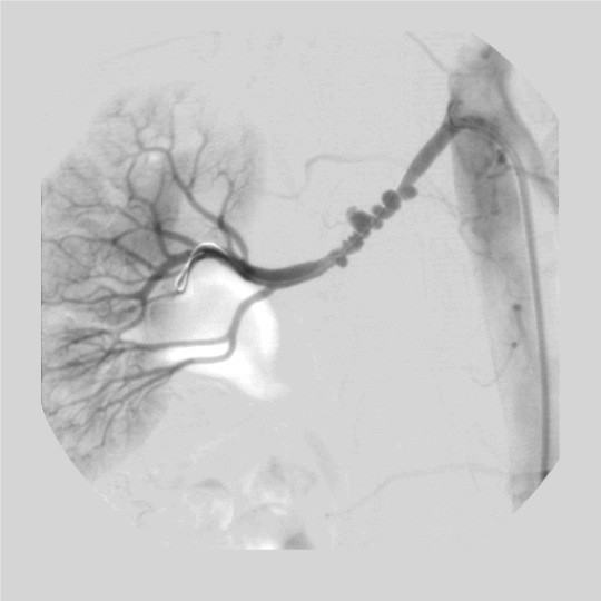
Image Quiz - Fibromuscular Disease
The answer:
Fibromuscular disease (FMD)
The Case
What's the diagnosis?

- Renal artery involvement ≈ 60% to 75% of cases and ≈35% bilateral
- Carotid or vertebral arteries involvement ≈ 25%-30% of cases.
- Intracranial involvement rare
- ≈ 28% of cases multiple arterial systems are involved- renal arteries are almost always involved.
- FMD classified histologically according to whether it predominately affects the arterial media, intima, or adventitia.
- Medial dysplasia
- 3 histologic types:
- Medial fibroplasia ≈ 75% to 80% of all cases of FMD; characteristic string of beads with diameters larger than the normal artery size in the middle and distal portion of the main renal arteries and its branches. On histologic cross-section of the artery, the media has both very thin and very thick areas without inflammatory cells. The thin areas may develop aneurysms in a minority of patients.
- Perimedial fibroplasia: less than 10% of cases of FMD. It tends to occur in girls between 5 and 15 years old. It may progress to renal failure if left untreated. Perimedial disease also resembles a string of beads but with fewer beads and with diameters smaller than the normal artery size. Collateral arteries often form around the area of stenosis. Histologically, there is extensive collagen deposition in the outer half of the media.
- Medial hyperplasia is rare. It appears angiographically as a concentric focal band and histologically as smooth muscle cell hyperplasia without fibrosis. Medial hyperplasia resembles intimal fibroplasia and may not be a distinct condition.
- Intimal fibroplasia: less than 10% of cases of FMD.
- Adventitial (periarterial) fibroplasia: less than1% of FMD.
The Case
What's the diagnosis?

Sunday, December 2, 2012
Friday, November 30, 2012
Electrolyte Quiz
The Answer: B
Hypercalcemia is a common complication of
malignancies. The etiology of hypercalcemia is enhanced bone resorption. This
reflects: release of parathyroid hormone (PTH)-related peptide (PTHrP), local
release of bone resorptive cytokines by lytic tumors, and production by the
tumor of 1,25 Vitamin D3.
Treatment comprises of: hydration, forced alkaline
diuresis, use of calcitonin, and administration of bisphosphonates. Bisphosphonates are effective for the
management of chronic hypercalcemia but are contraindicated in patients with
moderate to severe renal insufficiency. Even if one considered using a
bisphosphonate it would be after other strategies had been exhausted. Hydration
is key.
In this case, the patient appears to have been well hydrated (note the
absence of orthostatic changes, the normal heart rate, and the moist mucous
membranes). Hemodialysis is also an effective therapy for the treatment of
hypercalcemia but may not be appropriate as yet (i.e., as the first step).
The
next step in management should be to start calcitonin, which is effective over
a few hours, and mithramicin. Corticosteroid administration could also be
considered.
A 64-year old woman with a history of breast cancer is admitted to the hospital with confusion, nausea and vomiting. She was doing well until 1 week ago when she noticed a marked increase in her urine output. She has a past medical history of Her2+ breast cancer diagnosed 2 years previously and she is status post adjuvant chemotherapy, lymph node clearance left axilla, and radiation therapy. Her physical examination shows her to have a blood pressure of 118/70 mmHg with no orthostatic change and a heart rate of 82 beats per minute. Her mucous membranes are moist. Except for a left axillary wall scar and moderate hepatomegaly, the rest of the physical examination is unremarkable. Her serum calcium adjusted for her serum albumin is 14.2 mg/dL. Her other electrolytes are normal. Her BUN is 68 mg/dL and her serum creatinine is 5.2 mg/dL. She has been hydrated with 3 liters of 0.9% saline administered at 100 mL/hr. Her repeat serum calcium is 12.9 mg/dL and she remains confused. You are asked to provide additional recommendations regarding management.
The most appropriate next best step is to:
A.) Start the patient on a bisphosphonate
B.) Start this patient on calcitonin and mithramycin
C.) Continue the intravenous hydration
D.) No treatment
E.) Begin hemodialysis
Thursday, November 29, 2012
Electrolyte Quiz
A 64-year old woman with a
history of breast cancer is admitted to the hospital with
confusion, nausea and vomiting. She was doing well until 1 week ago when she
noticed a marked increase in her urine output. She has a past medical history of Her2+ breast cancer diagnosed 2 years previously
and she is status post adjuvant chemotherapy, lymph node clearance left axilla, and radiation therapy. Her physical
examination shows her to have a blood pressure of 118/70 mmHg with no
orthostatic change and a heart rate of 82 beats per minute. Her mucous
membranes are moist. Except for a left axillary wall scar and moderate
hepatomegaly, the rest of the physical examination is unremarkable. Her serum
calcium adjusted for her serum albumin is 14.2 mg/dL. Her other electrolytes
are normal. Her BUN is 68 mg/dL and her serum creatinine is 5.2 mg/dL. She has
been hydrated with 3 liters of 0.9% saline administered at 100 mL/hr. Her
repeat serum calcium is 12.9 mg/dL and she remains confused. You are asked to
provide additional recommendations regarding management.
The most appropriate next best step is to:
A.) Start the patient on a bisphosphonate
B.) Start this patient on calcitonin and mithramycin
C.) Continue the intravenous hydration
D.) No treatment
E.) Begin hemodialysis
Tuesday, November 27, 2012
Image Quiz - Staghorn calculus
Answer
The picture - a plain abdominal X ray - show a right sided staghorn calculus.
A staghorn calculus (also sometimes called coral calculi) is a very large calculus that nearly fills the calyceal system, with extensions into calyces that give the appearance of a stag's (deer) horns.
The formal definition of a stag horn calculus is an "upper urinary tract stone that involves the renal pelvis and extends into at least 2 calyces. All types of urinary stones can potentially form staghorn calculi, but approximately 75% are composed of a struvite-carbonate-apatite matrix. These so-called struvite stones, named after the 19th-century Russian diplomat Baron von Struve (friend and mentor to 19th-century discoverer Ulex), are also known as triple-phosphate (3 cations associated with 1 anion), infection (or infection-induced), phosphatic, and urease stones. (Ref: Meng et al emedicine)
Question
This is a plain X ray of the abdomen. What does it show?
Monday, November 26, 2012
Friday, November 23, 2012
Image Quiz
The answer
Secondary FSGS in a patient with unilateral renal agenesis.
The image shows:
Panel A: low power LM. Four enlarged glomeruli are present. One of them (focal), second from left, has a segmental area of capillary collapse, illustrated in greater detail in the next slide. Notice also a small area of tubules (center, upper half) with thickened basement membranes and slight interstitial fibrosis. (PAS stain).
Panel B: greater detail of segmental lesion. Notice collapse of capillaries between 2 and 10 o'clock, with hyalinosis, most marked at 2 and 5 o'clock. (PAS stain).
Panel C: EM details of the capillary walls. This EM shows slight mesangial expansion, mild thickening of the GBM, no deposits, no hyaline accumulation in these loops, and relatively well preserved foot processes. The "fusion" occurs in general much more focally in these secondary forms of FGS when compared to the more diffuse obliteration of foot processes in the primary forms.
The Case
This is a 43 year old patient with a serum creatinine of 1.8 mg/dL and 2 grams of albuminuria by UACR. The kidney biopsy is shown below. What's the diagnosis?
Secondary FSGS in a patient with unilateral renal agenesis.
The image shows:
Panel A: low power LM. Four enlarged glomeruli are present. One of them (focal), second from left, has a segmental area of capillary collapse, illustrated in greater detail in the next slide. Notice also a small area of tubules (center, upper half) with thickened basement membranes and slight interstitial fibrosis. (PAS stain).
Panel B: greater detail of segmental lesion. Notice collapse of capillaries between 2 and 10 o'clock, with hyalinosis, most marked at 2 and 5 o'clock. (PAS stain).
Panel C: EM details of the capillary walls. This EM shows slight mesangial expansion, mild thickening of the GBM, no deposits, no hyaline accumulation in these loops, and relatively well preserved foot processes. The "fusion" occurs in general much more focally in these secondary forms of FGS when compared to the more diffuse obliteration of foot processes in the primary forms.
The Case
This is a 43 year old patient with a serum creatinine of 1.8 mg/dL and 2 grams of albuminuria by UACR. The kidney biopsy is shown below. What's the diagnosis?
Thursday, November 22, 2012
Friday, November 16, 2012
Image Quiz
JE is a 61 year old white male with ESRD of unknown etiology who began chronic hemodialysis in August 1987. His past medical history is significant for Crohn's disease requiring 3 1/2 feet of bowel resection in 1980. He received a cadaveric renal allograft on 8/1/88. The initial post-transplant course was complicated by delayed graft function and his immunosuppressive regimen consisted of prednisone, azathioprine and anti-thymocyte globulin.
He was started on cyclosporine at a time when his urine output was increasing, however within four days he developed decreased urine output and the CsA was discontinued and he was maintained on azathioprine and prednisone.
An allograft biopsy was performed on the 22nd post-operative day and repeated on the 40th post-operative day due to continued poor allograft function. A third allograft biopsy was performed on the 65th post-transplant day.
Use the images below to make the diagnosis!
The renal biopsy is shown below:
![]() |
| Case and histopath courtesy of Dr. Helmut Rennke, BWH/HMS |
Wednesday, November 7, 2012
Image Quiz
The Answer
BK virus nephropathy (nephritis) or BKVN
Question
What's the diagnosis?
BK virus nephropathy (nephritis) or BKVN
Question
What's the diagnosis?
Explanation
Dall and Hariharan provide a fantastic review of BK virus nephritis (BKVN) in CJASN that is available open access.
Some take-aways from the paper:
1. The term “BK” originated from a patient's initials, in whom it was first detected in 1971
2. Prevalence of BK viremia within 1 yr after transplantation is approximately 20%
3. BKVN has increasingly been recognized as an early event and occurs within the first year after transplantation.
4. Quantification of viral load in blood and urine with either viral DNA or mRNA (to VP-1) has been used to diagnose BKVN.
5. Decoy cells seen on urine cytology have been observed in patients with BKVN and originate from infected renal tubular cells with nuclei altered by viral inclusions. The presence of decoy cells is a sensitive (100%) measure but has a low positive predictive value of 29% for the diagnosis of BKVN.
6. Pathologic findings of infection include viral cytopathic changes in the epithelium of tubules, glomeruli, and collecting ducts with interstitial inflammation and varying degrees of tubular atrophy or fibrosis (42). Patchy interstitial involvement is common early in the course of disease followed by extensive inflammation progressing to fibrosis.
Tuesday, November 6, 2012
Monday, October 22, 2012
Monday, October 1, 2012
Glomerulonephritis Quiz
The answer is - MPGN - C.
The patient has a low C3, azotemia and a nephritic urine and a normal ASLO titer and a normal ANA, anti-GBM and ANCA.
17-year old female comes to your office complaining of an episode of gross hematuria. For the past 5 days she has had pharyngitis, nasal discharge, and flu-like symptoms. Over the past two days has taken 8 Bactrim tablets left over from a previous episode of bronchitis. She reports no change in her urine output.
The patient has a low C3, azotemia and a nephritic urine and a normal ASLO titer and a normal ANA, anti-GBM and ANCA.
17-year old female comes to your office complaining of an episode of gross hematuria. For the past 5 days she has had pharyngitis, nasal discharge, and flu-like symptoms. Over the past two days has taken 8 Bactrim tablets left over from a previous episode of bronchitis. She reports no change in her urine output.
On examination her key findings: her weight is 55 kg, Pulse-76 bpm, BP 130/80 mmHg. Heart, lungs, abdomen are normal. There is no rash or edema.
Laboratory Studies
Hb-14 g/dL, WBC-5400 (normal diff), BUN-25, creat-1.7, C3 88 (100-233), C4-28 (14-48), ASLO-200 (normal is less than 200), ANA, GBM, ANCA negative. Urine protein2.4g/24h, UA-2+ protein, 20-30 RBCs, 4-6 WBC, occasional rbc casts
During close outpatient monitoring, blood pressure and creat remain unchanged. On follow-up 12 weeks later, laboratory studies reveal;
Bun-24, creat-1.9, C3-70, C4-20, UA- 2+protein, 3-8 RBCs, 0-4 wbcs, no casts
A renal biopsy is performed:
Which of the following is the most likely diagnosis?
A. Allergic interstitial nephritis
B. Post-strep glomerulonephritis
C. Membranoproliferative glomerulonephritis
D. IgA Nephropathy
(Source: Dr. Brad Denker, Renal Division, Beth Israel Deaconess Medical Center/Harvard Medical School)
Sunday, September 30, 2012
Glomerulonephritis Quiz
17-year old female comes to
your office complaining of an episode of gross hematuria. For the past 5 days
she has had pharyngitis, nasal discharge, and flu-like symptoms. Over the past
two days has taken 8 Bactrim tablets left over from a previous episode of
bronchitis. She reports no change in her urine output.
On examination her key findings: her weight is 55
kg, Pulse-76 bpm, BP 130/80 mmHg. Heart, lungs, abdomen are normal. There is no rash or edema.
Laboratory Studies
Hb-14 g/dL, WBC-5400 (normal diff), BUN-25, creat-1.7, C388 (100-233),
C4-28 (14-48), ASLO-200 (normal is less
than 200), ANA, GBM, ANCA negative. Urine protein2.4g/24h, UA-2+ protein, 20-30
RBCs, 4-6 WBC, occasional rbc casts
During close outpatient monitoring, blood pressure and creat
remain unchanged. On follow-up 12 weeks later, laboratory studies reveal;
Bun-24, creat-1.9, C3-70, C4-20, UA- 2+protein, 3-8 RBCs, 0-4 wbcs, no casts
A renal biopsy is performed:
Which of the following
is the most likely diagnosis?
A. Allergic interstitial nephritis
B. Post-strep glomerulonephritis
C. Membranoproliferative glomerulonephritis
D. IgA Nephropathy
(Source: Dr. Brad Denker, Renal Division, Beth Israel Deaconess Medical Center/Harvard Medical School)
Sunday, September 23, 2012
Sunday, September 9, 2012
Hypertension Quiz
Answer
B
The Question
A 20-year-old female is referred for work-up of newly diagnosed
hypertension.
Laboratory Studies
Serum sodium 144 mEq/L
Serum potassium 3.2 mEq/L
Serum chloride 107 mEq/L
Serum bicarbonate 29 mEq/L
Blood urea nitrogen 19 mg/dL
Serum creatinine 1 mg/dL
Plasma renin activity 0.8 ng/mL/hr (Normal range 1-6)
Plasma aldosterone 4.2 ng/dL (Normal range 5-20)
Which of the following tests would be appropriate to send next:
A. 24 hr urine aldosterone
B. 24 hr urine cortisol
C. 24 hr urine 18-hydroxycortisol and 18-oxocortisol
D. Doppler ultrasound of the renal arteries
E. Serum metanephrines
Explanation
This patient has secondary hypertension associated with
hyperaldosteronism but suppressed levels of both renin and aldosterone. The
differential diagnosis is Cushing syndrome, which could be diagnosed from the
24 hr urine cortisol, Liddle syndrome, glycyrrhizic acid intoxication and the
syndrome of apparent mineralocorticoid excess. Primary hype aldosteronism and
glucocorticoid-remediable aldosteronism (associated with elevated 24 hr urine
levels of aldosterone and 18-hydroxycortisol/18oxocortisol, respectively) would
be associated with elevated aldosterone levels. Renal artery stenosis,
diagnosable by Doppler ultrasound, would be associated with elevated renin and
aldosterone levels. Serum metanephrines are used in the diagnosis of
pheochromocytoma, which does not cause metabolic alkalosis.
(Source: Dr. Alan S. Yu, Professor of Medicine and Director, Kidney Institute, University of Kansas Medical Center, USA).